Breast Cancer
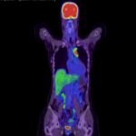
PET CT plays a significant role in the diagnosis, staging, treatment planning, and monitoring of breast cancer. Here are the key roles and benefits of PET CT in managing breast cancer:
1. Diagnosis and Detection
- Primary Tumor Identification: PET CT can help in detecting primary breast tumors, especially in cases where conventional imaging methods are inconclusive.
- Differentiation of Benign and Malignant Lesions: PET CT is valuable in differentiating between benign and malignant lesions, which can reduce unnecessary biopsies and surgeries.
2. Staging
- Locoregional Staging: PET CT can assess the extent of the primary tumor and involvement of regional lymph nodes. This is crucial for accurate staging and treatment planning.
- Distant Metastasis Detection: PET CT is highly effective in detecting distant metastases, including in bones, liver, lungs, and brain. This comprehensive staging helps in tailoring appropriate systemic therapy.
3. Treatment Planning
- Radiotherapy Planning: PET CT provides detailed anatomical and functional information that aids in precise radiotherapy planning, ensuring better targeting of the tumor while sparing healthy tissues.
- Surgical Planning: It can help surgeons understand the extent of the disease and plan the surgical approach, particularly in cases requiring extensive resections.
4. Monitoring Response to Therapy
- Chemotherapy and Radiotherapy Response: PET CT can evaluate the metabolic response of tumors to chemotherapy and radiotherapy. Changes in metabolic activity often precede changes in tumor size, allowing for early assessment of treatment efficacy.
- Neoadjuvant Therapy: For patients receiving neoadjuvant therapy (treatment before surgery), PET CT can assess the tumor’s response, which may influence the subsequent surgical approach.
5. Detection of Recurrence
- Surveillance: In the follow-up period, PET CT is useful in monitoring for disease recurrence. It can detect recurrences earlier than conventional imaging modalities by identifying metabolic changes.
- Evaluation of Suspicious Findings: When there are equivocal findings on mammography, ultrasound, or MRI, PET CT can provide additional information to clarify these findings.
6. Prognostic Value
- Prognostic Indicators: PET CT can provide prognostic information based on the metabolic activity of the tumor. Higher metabolic activity as indicated by standardized uptake values (SUVs) may correlate with more aggressive disease and poorer prognosis.
Limitations and Considerations
- False Positives and Negatives: PET CT can sometimes produce false-positive results due to inflammation or infection and false-negative results, particularly in small lesions or certain types of breast cancer with low metabolic activity.
- Cost and Accessibility: PET CT is more expensive and less accessible than conventional imaging techniques, which may limit its use in some settings.
- Radiation Exposure: While PET CT involves radiation exposure, the benefits often outweigh the risks, especially in the context of accurate staging and monitoring.
Overall, PET CT is a powerful tool in the multidisciplinary management of breast cancer, offering detailed insights that can significantly influence treatment decisions and improve patient outcomes