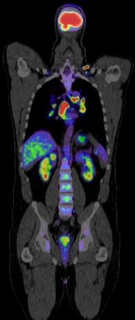
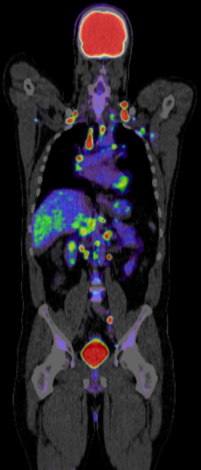
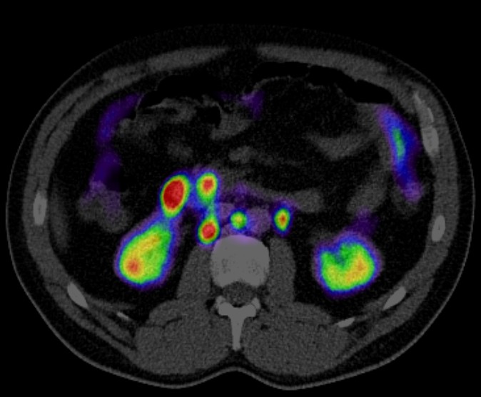
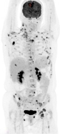
Gastrointestinal Stromal Tumour (GIST)


Positron emission tomography-computed tomography (PET CT) plays a significant role in the diagnosis, staging, and management of gastrointestinal stromal tumors (GISTs). Here are some key aspects of its role:
1. Diagnosis and Initial Staging
- Detection of Primary Tumor: PET CT is useful in detecting the primary GIST, especially when the tumor is metabolically active and uptakes the radiotracer, typically 18F-fluorodeoxyglucose (FDG).
- Assessing Tumor Extent: It helps in evaluating the extent of the disease by identifying primary and metastatic sites, providing a comprehensive view that can guide biopsy and treatment planning.
2. Assessment of Treatment Response
- Monitoring Response to Therapy: PET CT is particularly valuable in assessing the response to tyrosine kinase inhibitors (TKIs) like imatinib. Reduction in FDG uptake often precedes changes in tumor size, indicating metabolic response even before anatomical changes are evident.
- Early Prediction of Response: Early PET CT scans post-therapy can predict the effectiveness of treatment, allowing for timely modifications in therapeutic strategy.
3. Detection of Recurrence
- Surveillance: PET CT is used in the follow-up of patients to detect recurrence. Since GISTs can recur even after complete surgical resection, regular monitoring is crucial.
- Differentiation of Recurrence from Scar Tissue: PET CT helps differentiate between metabolically active recurrent tumor and non-metabolic post-surgical scar tissue.
4. Prognostic Value
- Baseline Metabolic Activity: High baseline FDG uptake in GISTs is often associated with more aggressive disease and poorer prognosis. PET CT helps in identifying high-risk patients who might benefit from more aggressive or prolonged therapy.
5. Guiding Biopsy
- Targeting Biopsy Sites: By identifying the most metabolically active and thus most representative areas of the tumor, PET CT can guide biopsy to obtain the most diagnostically useful tissue samples.
6. Evaluation of Metastasis
- Whole-body Imaging: PET CT provides whole-body imaging, making it superior in detecting metastatic spread to distant organs compared to other imaging modalities that may focus only on certain regions.
PET CT is an invaluable tool in the management of GISTs, providing critical information from initial diagnosis through treatment and follow-up. Its ability to assess metabolic activity offers advantages over purely anatomical imaging techniques, enabling early detection of treatment response, accurate staging, and identification of recurrences. As such, PET CT contributes to more personalized and effective management strategies for patients with GISTs.
