Gynaecological Cancer


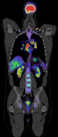
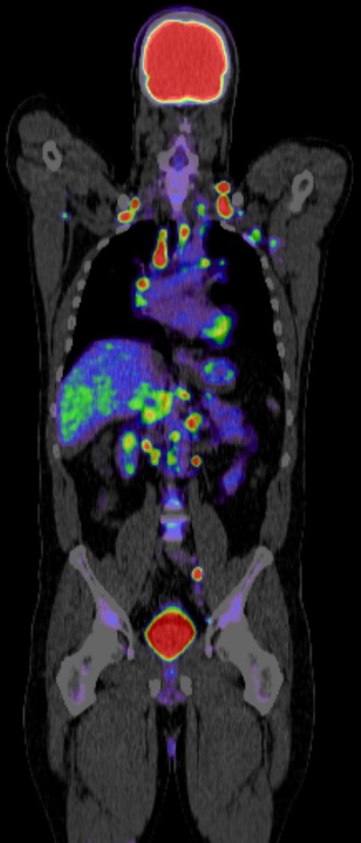
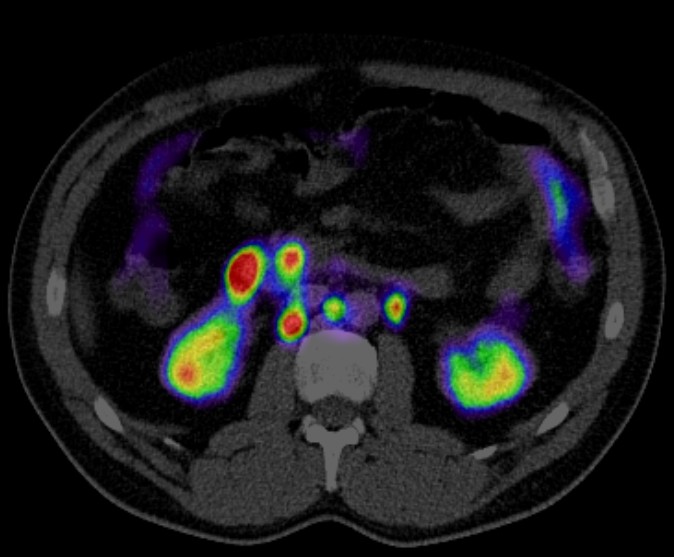
PET CT (Positron Emission Tomography – Computed Tomography) has become an important tool in the management of gynecological cancers. It combines the functional imaging of PET with the anatomical imaging of CT, providing detailed information about both the structure and function of tissues. Here are key roles PET CT plays in gynecological cancers:
1. Diagnosis and Staging
- Detection of Primary Tumors: PET CT can help in identifying primary gynecological tumors, particularly in cancers of the cervix, ovary, and endometrium.
- Staging: It provides detailed information on the extent of the disease, including lymph node involvement and distant metastasis. Accurate staging is crucial for determining the appropriate treatment strategy.
2. Treatment Planning
- Guiding Biopsies: PET CT can help in targeting the most metabolically active areas of a tumor for biopsy, ensuring a more accurate diagnosis.
- Radiation Therapy Planning: It aids in delineating the tumor boundaries more precisely, which is essential for planning effective radiation therapy while sparing healthy tissues.
3. Assessment of Treatment Response
- Monitoring Response: PET CT is used to monitor the metabolic response of tumors to treatment (chemotherapy, radiation therapy, or surgery). A decrease in metabolic activity on PET CT usually indicates a good response to treatment.
- Early Detection of Recurrence: It can detect metabolic changes that precede anatomical changes, allowing for the early detection of recurrent disease.
4. Prognostic Value
- Predicting Outcomes: The metabolic activity of tumors as seen on PET CT can be predictive of patient outcomes. Higher uptake of the radiotracer often correlates with more aggressive disease and a poorer prognosis.
5. Detection of Occult Disease
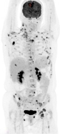
- Identifying Metastases: PET CT is particularly useful in identifying occult metastases that may not be detected by other imaging modalities. This is important for ensuring comprehensive treatment planning.
Specific Gynecological Cancers
Cervical Cancer
- PET CT is used for initial staging, particularly in detecting lymph node involvement and distant metastases.
- It is useful for restaging after treatment and for detecting recurrences.
Ovarian Cancer
- While CT and MRI are commonly used, PET CT can be beneficial in cases where there is a suspicion of recurrent disease, especially when other imaging modalities are inconclusive.
- It helps in detecting small peritoneal implants and lymph node metastases.
Endometrial Cancer
- PET CT is used for initial staging, especially in high-risk patients, to assess lymph node and distant metastasis.
- It is also useful in the follow-up of patients with suspected recurrence.
Advantages and Limitations
Advantages:
- High Sensitivity and Specificity: PET CT provides high sensitivity and specificity for detecting both primary and metastatic lesions.
- Whole-body Imaging: It allows for a comprehensive evaluation of the entire body in a single scan.
- Functional Imaging: PET CT provides metabolic information that can be more indicative of malignancy than structural changes alone.
Limitations:
- False Positives/Negatives: Inflammation and infection can cause false positives, while small lesions may be missed.
- Radiation Exposure: PET CT involves exposure to ionizing radiation, which is a consideration, especially in younger patients.
- Cost: It is more expensive compared to other imaging modalities like CT or MRI.
PET CT plays a critical role in the diagnosis, staging, treatment planning, and follow-up of gynecological cancers. Its ability to provide both functional and anatomical information makes it a valuable tool in the comprehensive management of these cancers. However, it should be used judiciously, considering its limitations and the clinical context.
