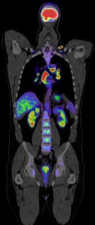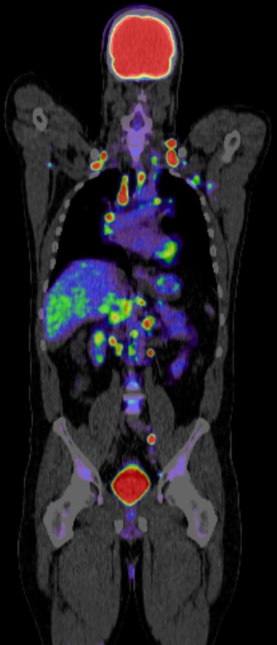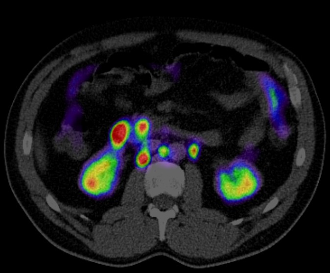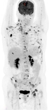


PET CT (positron emission tomography-computed tomography) plays a crucial role in the diagnosis, staging, and monitoring of multiple myeloma. Here’s how:
Diagnosis: PET CT can detect bone lesions earlier and more accurately than conventional imaging techniques like X-rays. It helps in identifying active myeloma lesions throughout the skeleton, even in areas where bone density is not significantly compromised. This is important because myeloma can affect multiple bones in the body.
Staging: PET CT assists in determining the extent of myeloma involvement in bones and other tissues. By identifying the spread and activity of the disease, it aids in staging, which is crucial for determining the appropriate treatment strategy.
Response Assessment: PET CT is valuable in assessing the response to treatment. It can detect changes in metabolic activity and the size of lesions, allowing doctors to gauge the effectiveness of therapies. Decreased metabolic activity or reduction in the size of lesions indicates a favorable response to treatment, while increased activity may suggest disease progression or resistance to treatment.
Monitoring: After treatment initiation, regular PET CT scans can help in monitoring the disease progression or remission. This monitoring is crucial for adjusting treatment plans as needed, such as changing medications or pursuing additional therapies if there’s evidence of disease relapse or progression.
Prognostic Information: PET CT findings, such as the extent and intensity of bone lesions, can provide prognostic information. Higher metabolic activity and extensive bone involvement may indicate a poorer prognosis, whereas a good response to treatment on PET CT scans may suggest a better overall outlook.
In summary, PET CT is an essential imaging modality in the management of multiple myeloma, providing valuable diagnostic, staging, monitoring, and prognostic information that helps guide treatment decisions and improve patient outcomes.
