Pancreatic Carcinoma


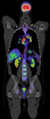
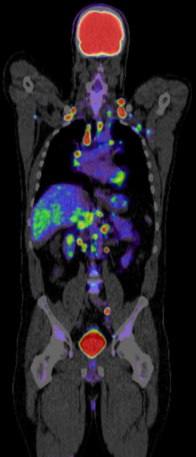
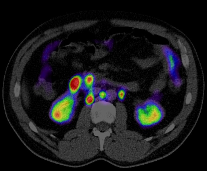
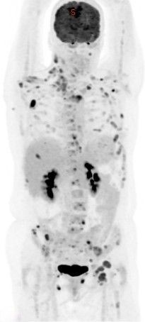
PET CT is a powerful tool used in the diagnosis, staging, treatment and monitoring of pancreatic carcinoma.
Diagnosis
Detection: PET/CT can detect primary pancreatic tumors by highlighting areas of increased metabolic activity typically associated with cancer cells.
Differentiation: It helps in differentiating benign from malignant pancreatic lesions, although it is not always definitive due to overlapping metabolic activity in inflammation and cancer.
Staging
Local and Distant Spread: PET CT is effective in evaluating the extent of the disease, including local invasion and distant metastases. This is crucial for planning surgery and other treatments.
Lymph Node Involvement: The technique is useful in assessing lymph node involvement, which is an important factor in staging and prognosis.
Treatment Planning
Guiding Surgery and Radiotherapy: PET CT helps in precise localization of the tumor, aiding in surgical planning and targeting radiation therapy.
Neoadjuvant Therapy Assessment: It can assess the response to neoadjuvant therapy (treatment given before the main treatment), helping to modify treatment plans based on the tumor’s metabolic response.
Monitoring and Follow-up
Treatment Response: PET/CT is valuable in monitoring the effectiveness of treatments such as chemotherapy, radiotherapy, or targeted therapies.
Detection of Recurrence: It can detect recurrence earlier than conventional imaging methods by identifying metabolic changes before structural changes become apparent.
Prognostic Value
Prognostic Biomarkers: The level of uptake of the radiotracer (commonly FDG, or fluorodeoxyglucose) can provide prognostic information. Higher uptake often correlates with more aggressive disease and poorer prognosis.
Limitations
False Positives/Negatives: Inflammation and infection can cause false positives due to increased FDG uptake, while small lesions may result in false negatives.
