Sarcoma


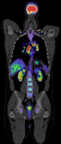
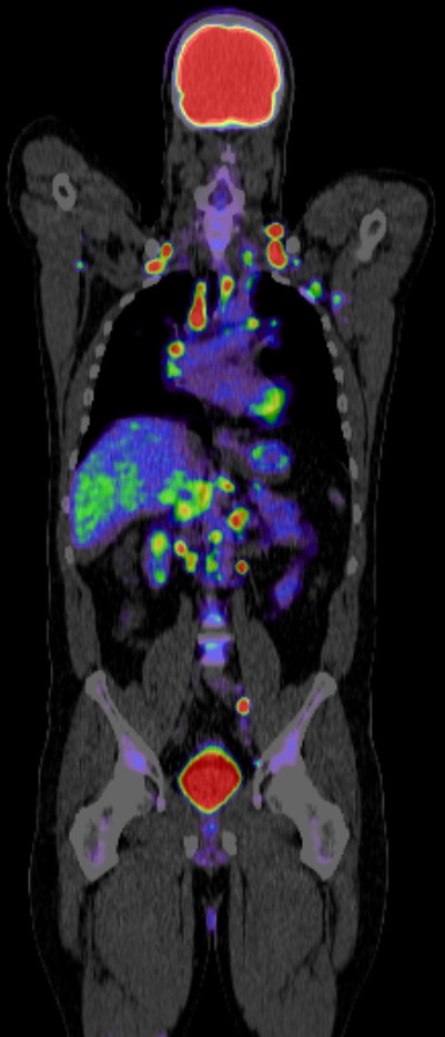
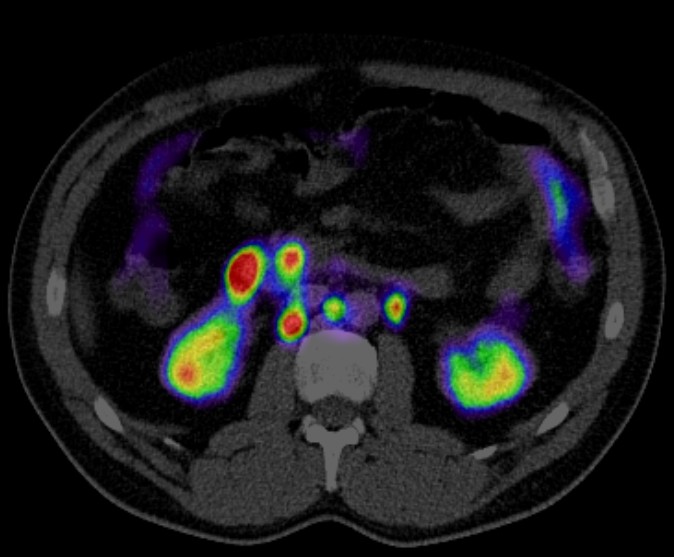
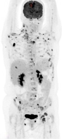
Positron Emission Tomography combined with Computed Tomography (PET CT) plays a significant role in the diagnosis, staging, treatment planning, and monitoring of sarcoma. Here’s a detailed look at its roles:
1. Diagnosis and Initial Staging
- Differentiation of Benign and Malignant Tumors: PET CT can help differentiate between benign and malignant lesions, which is crucial for accurate diagnosis.
- Extent of Disease: It provides comprehensive information about the primary tumor and any potential metastatic disease, which is essential for staging.
- Biopsy Guidance: PET CT can help guide biopsies to the most metabolically active areas of a tumor, ensuring that the sample taken is representative of the most aggressive part of the tumor.
2. Treatment Planning
- Radiation Therapy: PET CT helps in delineating the tumor boundaries more accurately, allowing for precise targeting in radiation therapy, thus sparing healthy tissue.
- Surgical Planning: The detailed imaging assists surgeons in planning the extent of surgical resection needed to remove the tumor completely.
3. Monitoring Response to Therapy
- Chemotherapy and Radiotherapy: PET CT is used to assess how well a tumor is responding to chemotherapy or radiotherapy by measuring changes in metabolic activity.
- Early Detection of Recurrence: It can detect recurrence earlier than conventional imaging by identifying metabolic changes before structural changes become apparent.
4. Prognostication
- Prognostic Value: The metabolic activity observed on PET CT scans can provide prognostic information. Higher uptake of the radiotracer often correlates with more aggressive disease and poorer prognosis.
5. Post-Treatment Surveillance
- Follow-Up: Regular PET CT scans are used during follow-up to monitor for recurrence and to assess the status of treated areas.
- Differentiation of Scar Tissue vs. Recurrence: PET CT helps distinguish between scar tissue from previous treatments and active tumor recurrence.
Technical Aspects and Radiotracers
- 18F-FDG (Fluorodeoxyglucose): The most commonly used radiotracer in PET/CT for sarcoma, as it highlights areas of high glucose metabolism typical of cancer cells.
- Other Tracers: In some cases, other radiotracers like 18F-FLT (fluorothymidine) might be used to provide additional information about tumor proliferation.
Limitations
- False Positives/Negatives: Inflammatory processes can also show high FDG uptake, leading to false positives. Similarly, low-grade tumors may not show significant FDG uptake, leading to false negatives.
- Cost and Accessibility: PET CT is a costly procedure and may not be available in all medical facilities.
Conclusion
PET CT is a valuable tool in the management of sarcoma, providing critical information that influences diagnosis, staging, treatment planning, response monitoring, and follow-up. Its ability to visualize metabolic activity offers advantages over traditional imaging methods, making it an integral part of modern oncological practice.
