Vasculitis


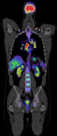
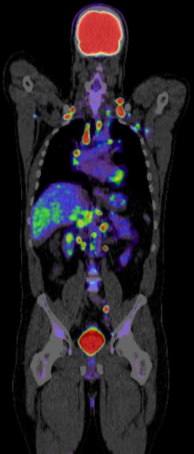
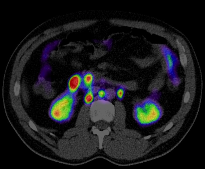
Positron Emission Tomography-Computed Tomography (PET-CT) plays a significant role in the diagnosis, assessment, and management of vasculitis, a group of disorders characterized by inflammation of blood vessels. Here are the key aspects of its role:
1. Diagnosis:
- Early Detection: PET CT can detect vascular inflammation at an early stage before structural changes become evident on other imaging modalities like MRI or CT alone.
- Identifying Active Disease: PET CT helps in distinguishing active inflammation from chronic fibrotic changes, which is crucial for determining the need for immunosuppressive therapy.
2. Assessment of Disease Extent:
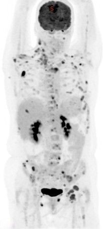
- Whole-Body Imaging: PET CT provides a comprehensive overview of the entire body, helping to identify the extent and distribution of vascular involvement. This is particularly useful in conditions like Takayasu arteritis and giant cell arteritis.
- Extra-Vascular Involvement: It can also detect inflammatory activity in tissues other than blood vessels, providing a complete assessment of the disease.
3. Evaluation of Disease Activity:
- Quantitative Measurement: PET CT can measure the degree of metabolic activity using standardized uptake values (SUVs), providing a quantitative assessment of inflammation.
- Monitoring Response to Treatment: By comparing PET CT scans before and after treatment, clinicians can evaluate the effectiveness of therapy and adjust treatment plans accordingly.
4. Differentiation from Other Conditions:
- Differentiation from Atherosclerosis: PET CT can help distinguish vasculitis from atherosclerotic disease, which is important since both conditions can present similarly on other imaging modalities but require different treatments.
- Detection of Infectious or Malignant Processes: It helps in ruling out infectious or malignant processes that can mimic vasculitis.
5. Guiding Biopsy:
- Targeted Biopsy: PET CT can identify the most active sites of inflammation, guiding biopsies to areas with the highest likelihood of yielding a definitive diagnosis, which is particularly useful in large-vessel vasculitis.
Specific Types of Vasculitis:
- Large Vessel Vasculitis (e.g., Takayasu Arteritis, Giant Cell Arteritis): PET CT is especially valuable in detecting inflammation in large vessels, where it can reveal circumferential uptake indicating active vasculitis.
- Medium and Small Vessel Vasculitis: While PET CT is less commonly used for these types, it can still provide valuable information in complex cases or when other modalities are inconclusive.
Limitations:
- Radiation Exposure: Repeated PET CT scans result in cumulative radiation exposure, which is a consideration, particularly in younger patients.
- Cost and Availability: PET CT is expensive and may not be readily available in all healthcare settings.
- False Positives/Negatives: Inflammation from other causes can result in false positives, and small-vessel inflammation may not be detectable on PET CT.
PET-CT is a powerful tool in the management of vasculitis, offering early detection, comprehensive assessment, and precise monitoring of disease activity. Its ability to provide functional and anatomical information makes it superior to many other imaging modalities in specific scenarios. However, its use should be balanced against considerations of cost, availability, and radiation exposure.
