Thymic Tumours


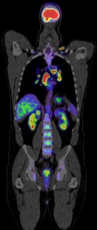
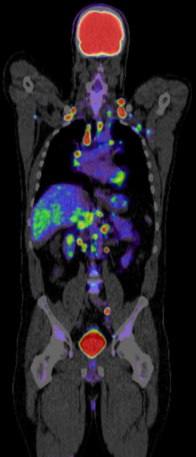
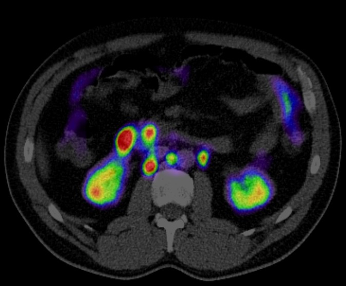
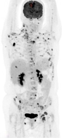
Positron Emission Tomography-Computed Tomography (PET CT) plays a significant role in the evaluation and management of thymic tumors. Here are some key aspects:
Staging: PET CT is crucial in determining the extent of the disease and staging thymic tumors. It helps in identifying whether the tumor is confined to the thymus or if it has spread to nearby structures such as the lungs, pleura, or lymph nodes.
Assessment of Metastasis: PET CT is effective in detecting distant metastases, particularly in the lungs, bones, and other distant organs. This information is vital for determining the appropriate treatment approach.
Differentiation of Tumor Types: PET CT can aid in distinguishing between thymomas and thymic carcinomas based on their metabolic activity. Thymomas typically exhibit lower metabolic activity compared to thymic carcinomas, which helps in guiding treatment decisions.
Evaluation of Treatment Response: PET CT is valuable for assessing the response to treatment, whether it be surgery, chemotherapy, radiation therapy, or a combination of these modalities. Changes in metabolic activity observed on PET CT scans can indicate the effectiveness of treatment and guide further management.
Detection of Recurrence: Following treatment, PET CT is useful for detecting recurrence or residual disease. It can identify areas of increased metabolic activity suggestive of recurrent disease, allowing for timely intervention.
Treatment Planning: PET CT findings contribute to treatment planning by providing information on the location and extent of the disease, helping in determining the need for further interventions such as surgery, radiation therapy, or systemic therapy.
Overall, PET CT is an important imaging modality in the comprehensive management of thymic tumors, assisting in diagnosis, staging, treatment planning, and monitoring of response to therapy.
